
Before & After
Gums around the canine tooth
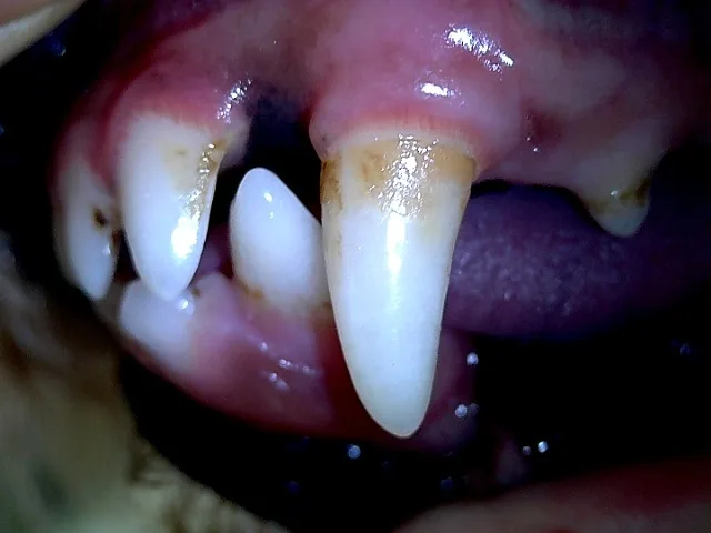
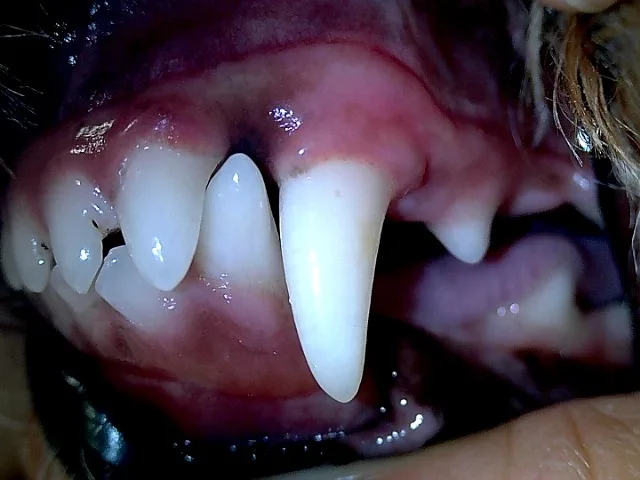
Left: tartar and staining around the canine / Right: tartar gone and gums settled
Key Summary
- Patient: 9-year-old Poodle, severe bad breath
- Diagnosis: Suspected tooth-root infection
- Treatment: Extraction and foreign object removal
- Result: Settled gums at 1 week
What happens when pus builds up at a dog's tooth root?
When bacteria infect the tissue around the tip of a tooth root and pus collects, it is called a tooth-root abscess. It develops when the nerve and blood vessels inside the tooth are damaged or infected and the inflammation spreads to the root tip.
The most common cause is periodontal disease: tartar and plaque build up, gum inflammation worsens, and the infection moves down toward the root. Untreated tooth damage or wear can also lead to it.
Early on there may be few signs, but as it progresses you may notice:
- Swelling on one side of the face, especially below the eye
- Worsening bad breath
- Pain when a certain area is touched, or trouble chewing
- In severe cases, a draining hole in the skin
Why did Mini come in?
| Patient | Mini (alias), 9-year-old female Poodle |
|---|---|
| Signs | A mild hacking cough and labored-looking breathing for about a week, and increasingly bad breath |
| History | Since early this year, the area under the left eye repeatedly puffed up like a water pouch and settled with medication at another clinic. Recently she had been chewing kibble carefully |
How did we look for the cause?
We started with the cough. Because heartworm prevention had not been given, we ran a heartworm test, which was negative.
The oral exam showed moderate tartar and bad breath. Together with the history of repeated swelling under the eye and discomfort when chewing, we first suspected a tooth-root infection.
Ongoing mouth pain and inflammation can increase swallowing or irritate the upper airway, so a cough can appear alongside. Also, if an infected root remains, medication alone tends to fail, so the affected tooth must be identified and removed.
We first calmed the cough with about a week of nebulizer treatment and medication, then planned dental X-rays under anesthesia and extraction if needed.
What tests were done before anesthesia?
The cough eased after medication, and we ran blood work to confirm she could be anesthetized safely. No abnormalities were found.
| Test | Result | Reference range | Meaning |
|---|---|---|---|
| BUN | 14.9 mg/dL | 9.2–29.2 | Normal |
| CREA | 0.4 mg/dL | 0.4–1.4 | Normal |
| PHOS | 4 mg/dL | 1.9–5 | Normal |
| GLU | 107 mg/dL | 75–128 | Normal |
| ALT | 36 U/L | 17–78 | Normal |
| ALB | 3.6 g/dL | 2.6–4.0 | Normal |
| ALKP | 96 U/L | 47–254 | Normal |
| TP | 6.8 g/dL | 5–7.2 | Normal |
| AST | 26 U/L | 17–44 | Normal |
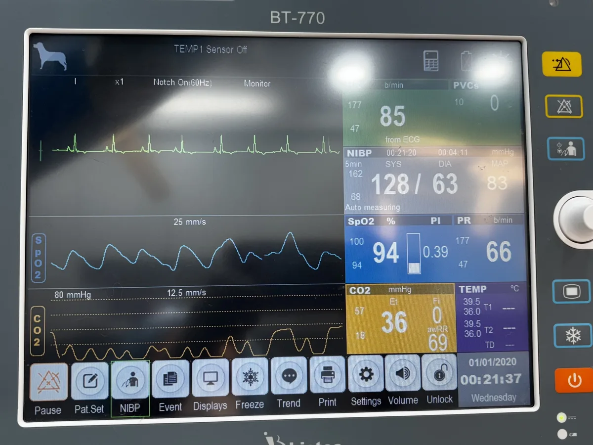
During anesthesia we continuously monitored heart rhythm, blood pressure, oxygen saturation and breathing.
What did the mouth and X-rays show?
Under anesthesia we examined the mouth closely. Tartar had built up, teeth were stained, and the gums were red.
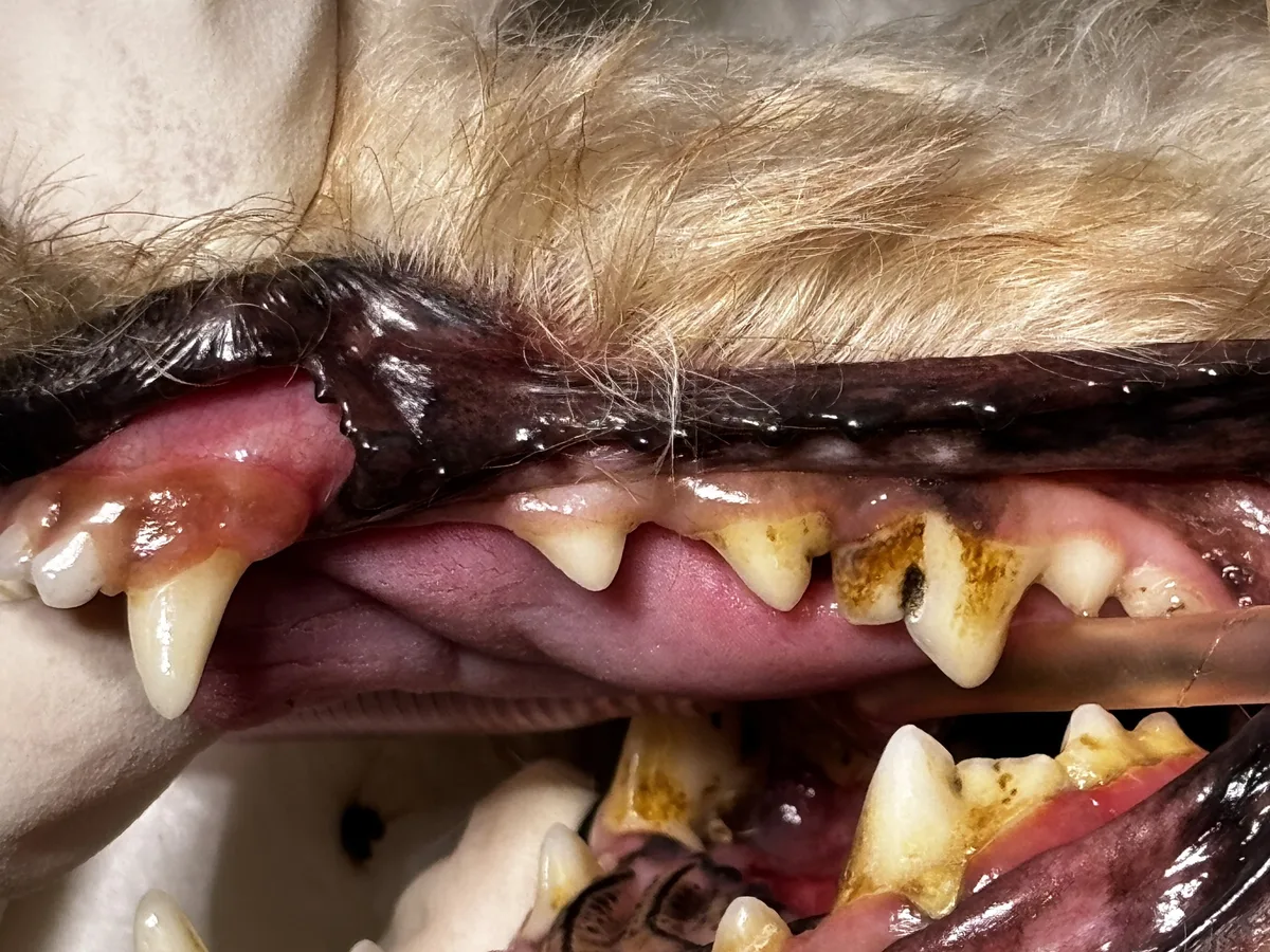
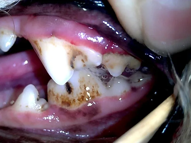
We also took chest X-rays under anesthesia, followed by dental X-rays to evaluate the tooth roots.
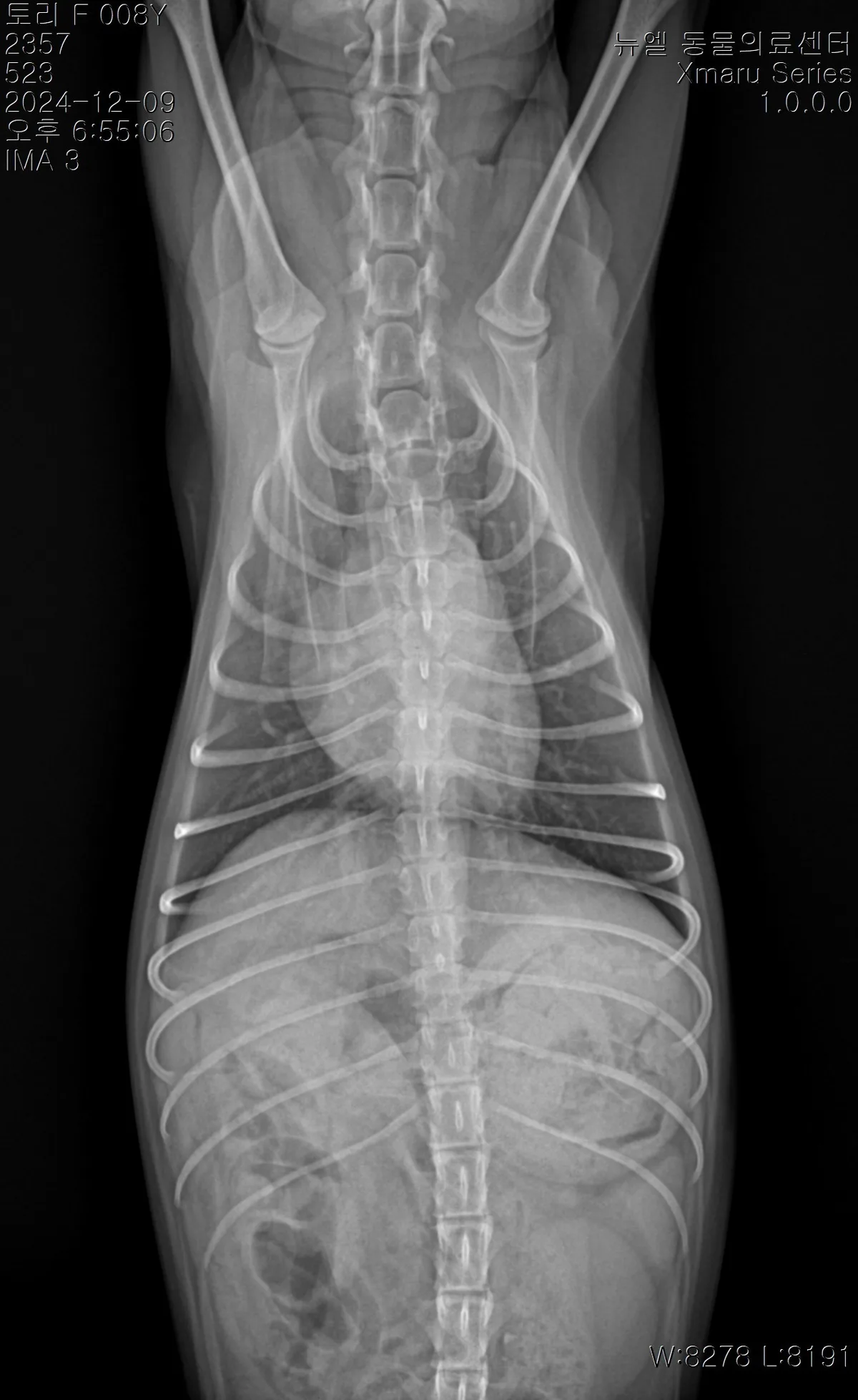
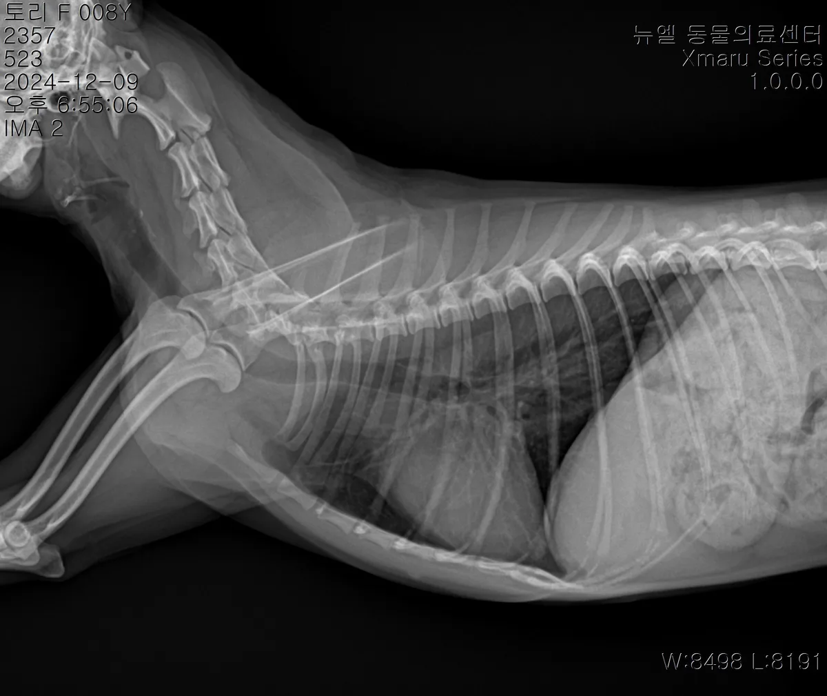
Left: chest, front view / Right: chest, side view
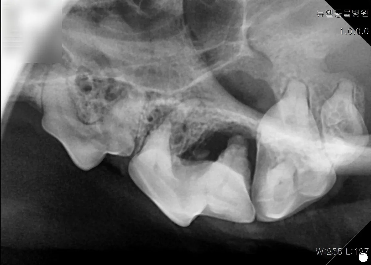
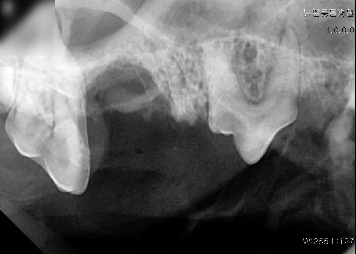
Dental X-rays showing the teeth and surrounding bone
How was she treated?
1. Extraction and foreign object removal
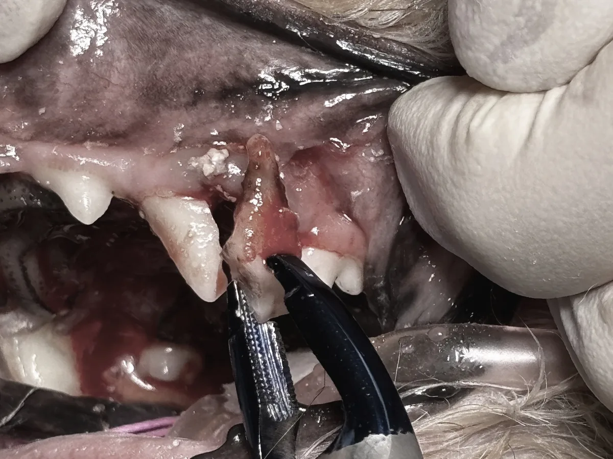
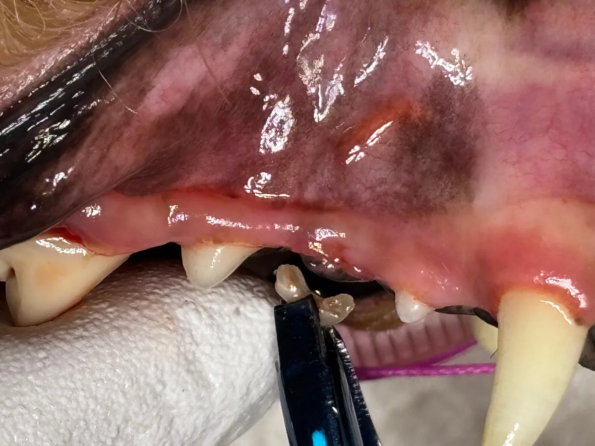
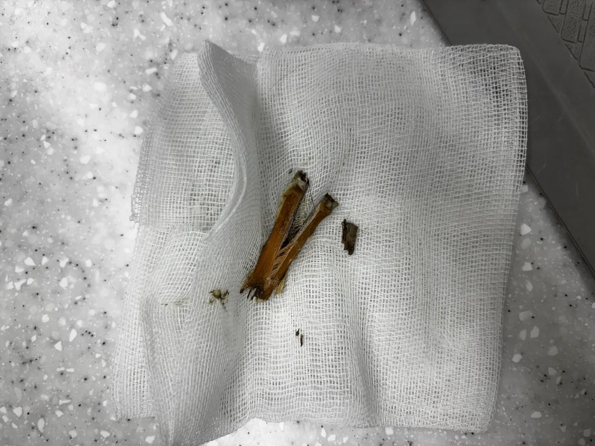
Based on the X-rays, we extracted the problem tooth and also removed a foreign object from the mouth. Not leaving an infected root behind is key to reducing recurrence.
⚠️ This post contains photos of a tooth extraction. They are hidden and shown only if you choose to open them.
⚠️ Extraction photos — tooth removal under anesthesia. Tap to view
2. Scaling and polishing
We removed the remaining tartar and polished the tooth surfaces so plaque does not stick as easily.
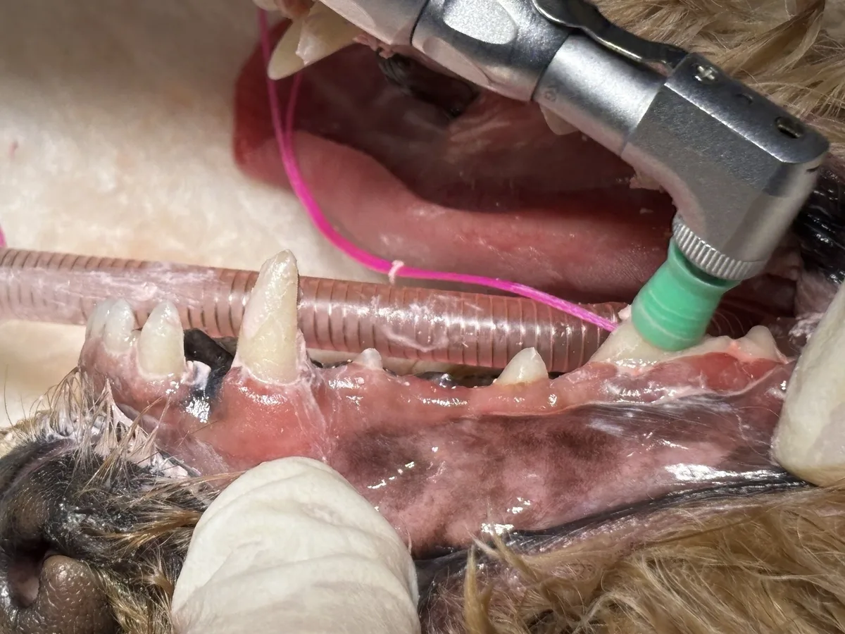
Before & after
Gums around the canine tooth
Left: before / Right: tidy gums one week later
Molar area
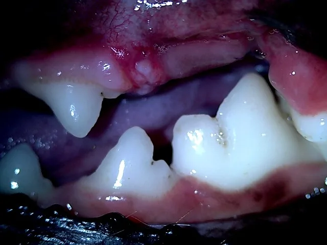
Left: thick tartar / Right: tartar gone and the extraction site healing
One week later the tartar was gone and the gums around the extraction site were settling well.
How should this be managed at home?
- Watch breath and chewing: check she chews comfortably and not on one side only.
- Check for facial swelling: look for swelling under the eye or on the cheek.
- Brushing and regular dental checks: managing tartar before it builds up is the best prevention.
Recovery varies from pet to pet.
Director's Comment
If swelling under the eye keeps coming back and settles with medication only to return, a tooth-root infection may be the cause. Medication alone tends to fail, so finding and removing the affected tooth is the key. In Mini's case a cough made it look like mouth and airway problems were mixed, so we first checked heartworm and respiratory status, then proceeded with treatment under safe anesthesia.
If bad breath worsens or your dog chews cautiously, we recommend a dental exam.
This case is also featured on Nuel Animal Medical Center's Naver blog.